MRI, EMG, X-Ray and Imaging Center
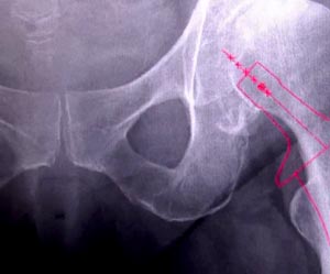
X-Ray
An X-ray is a painless process that uses low dose high-energy waves to take pictures of bones. X-rays can be used to evaluate bones, alignment, and joints, and they are utilized to help diagnose fractures, bone infections, tumors, arthritis, healing, and spatial relationships. During an X-ray, you will be asked to lie very still on a table and hold certain positions for a short period of time while images are taken. At Minneapolis Orthopaedics, we utilize state of the art low energy digital imaging processors that produce the highest quality images possible with the least radiation exposure.
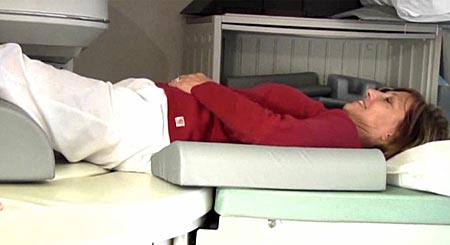
MRI (Magnetic Resonance Imaging) Scan
Unlike an X-ray, an MRI scan does not use radiation. By exerting a magnetic field, the MRI creates computer-generated images. The MRI is able to cut through multiple layers of tissues and show abnormalities in bone and soft tissues such as nerves, tendons, ligaments, cartilage, and muscles. MRI can also be used to verify loss of water in a disc and cartilage, joint abnormalities, narrowing or stenosis or disk herniation. During an MRI test, you will lie on a table for approximately 40 minutes. The machine's scanner then takes many pictures that are monitored by a technician and recorded. The MRI Scanner at Minneapolis Orthpaedics is a low energy high resolution open sided scanner that produces extemely detailed images. Our equipment passes the strictest standards and is ICAMRL (Intersocietal Commission for the Accreditation of Magnetic Resonance Laboratories) certified. The MRI Imaging Equipment employed at Minneapolis Orthopaedics is easily tolerated by people who are uncomfortable in closed spaces.
EMG (Electrodiagnostic Study)
An electromyogram (elec-tro-my-oh-gram) (EMG) is a test that looks at the function of the nerve roots that leave the spine and travel to muscles in the arms and legs. The test is performed by inserting tiny electrodes into the muscles of the lower extremity or by applying surface pads over the muscles. By looking for abnormal electrical signals in the muscles, the EMG can show whether a nerve is being irritated or pinched anywhere along its path. If the velocity or amplitude of the nerve transmission is reduced, then there may be pressure on the nerve along its course to the muscle and this is measured by the EMG test. This test can often localize where the pressure on the nerve exists which helps determine the best treatment for the problem. At Minneapolis Orthopaedics, we use a painless surface pad to measure nerve impulses combined with computer interpretion to yield the most accurate and pain free information about the function of your nerves.